Overview
Western blotting is a widely used laboratory method for analyzing proteins in a sample. It allows researchers to identify specific proteins, measure their abundance, and even detect changes in their structure. In cancer research, western blotting is a key tool for understanding how proteins contribute to tumor development, progression, and response to treatment.
Steps involved in Western Blotting
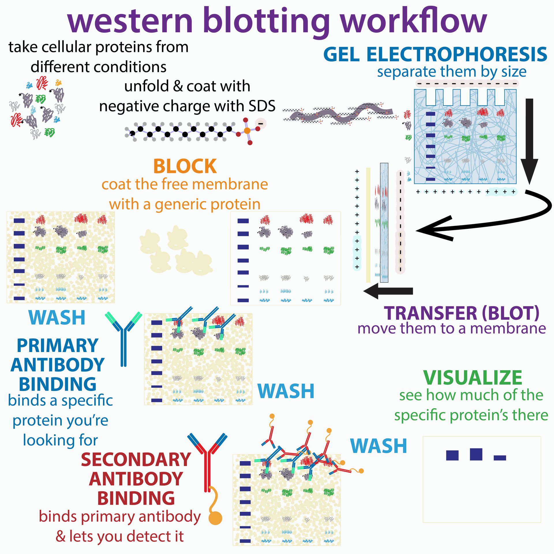
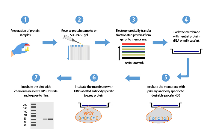
- Protein extraction: Proteins are isolated from the sample, typically tissue or cells, using specific buffers.
- Protein separation: Separates proteins based on size using a technique called SDS-PAGE. Proteins are treated with a detergent (SDS) to unfold them and then separated by an electric current through a gel.
- Protein transfer: Proteins are transferred from the gel to a membrane.
- Blocking: The membrane is treated with a solution to prevent irrelevant molecules from sticking to it.
- Antibody detection: A specific antibody designed to recognize the protein of interest is added to the membrane. If the protein is present, the antibody will bind to it.
- Signal generation: A second antibody, linked to a signaling molecule, is used to locate the first antibody. The signaling molecule allows the protein to be visualized.
- Analysis: The signal from the protein of interest is measured to determine its relative abundance in the sample.

Applications of Western Blotting in Cancer Research
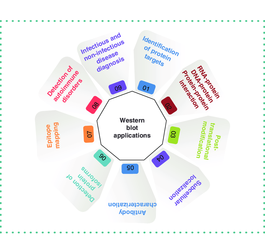
- Finding protein biomarkers: Western blotting can help identify proteins whose presence or absence indicates cancer.
- Studying protein modifications: Proteins can be chemically altered after they are made. Western blotting can be used to detect these modifications, which may be linked to cancer.
- Testing targeted therapies: Western blotting can be used to see if a cancer treatment affects the levels or modifications of specific proteins.

Purified 5HT1A Positive Control: Ensuring Accuracy in Western Blots:
- Antibody specificity: If the primary antibody used in the experiment is specific for the 5HT1A receptor, it should bind only to the purified protein in the positive control lane and generate a band at the expected size on the western blot.
- Transfer efficiency: The positive control ensures that proteins were efficiently transferred from the gel to the membrane during the blotting step. A strong band in the positive control lane indicates successful transfer.
- Western blot protocol functionality: The positive control serves as an internal control to verify that all steps of the western blot protocol, from protein extraction to detection, were performed correctly.
Western blotting experiments rely on specific proteins acting as controls to ensure the accuracy and validity of the results. One such control is a purified 5HT1A positive control. The 5HT1A receptor is a protein involved in the serotonin signaling pathway, and researchers frequently study its expression in various tissues.
In a western blot targeting the 5HT1A receptor, a purified 5HT1A positive control lane is included on the membrane alongside the experimental samples. This positive control is a commercially available preparation of purified 5HT1A protein at a known concentration. By running the positive control alongside the samples, researchers can confirm several aspects of their western blot:
Therefore, including a purified 5HT1A positive control strengthens the reliability of a western blot experiment investigating 5HT1A receptor expression.
Limitations of Western Blotting
- Relies on high-quality antibodies to work correctly.
- Provides an estimate, not an exact measurement, of protein abundance.
Conclusion
Western blotting is a fundamental tool in cancer research because it provides a relatively simple way to analyze protein expression. By revealing protein patterns and modifications, western blotting offers valuable insights into cancer biology, aiding in biomarker discovery, target identification, and drug development.
For a more in-depth explanation, please refer to the video linked below: